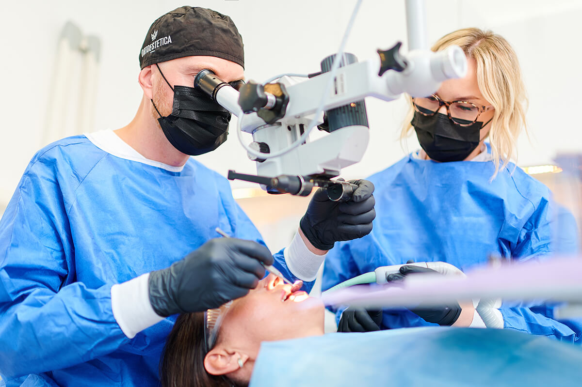
Microscope–Assisted Root Canal Therapy
Teeth decay is a contagious disease caused by bacteria whose multiplication is facilitated by the deposit which stores away upon the surface of the teeth and food particles that may get stuck in dental gaps. Appropriate oral hygiene allows to stop the development of the disease, which unfortunately is caused largely by the food that is eaten nowadays; the food is rich in sugar which is a perfect substrate for the bacteria, and it makes their multiplication a lot easier. Apart from the hygiene, it is important to remember about the check-up appointments with the dentist as these allow spotting early developmental stage of the disease when it is still relatively easy to cure it.
Every tooth has its nerve and blood vessels. If the developing decay does not get stopped and removed in time, and the tooth is not supplied with a filling, this situation will cause the bacteria to infect its nerve and blood vessels. In such a situation, the only solution which saves the tooth from extraction is root canal treatment. The entire root canal system of the tooth is very complex, which is why the microscope, precise disinfection, and accurate as well as hermetic filling of the canal are so crucially important to the endodontic treatment. The root canal treatment provided at our surgery is completed within a single visit. This kind of procedure is also known as endodontic treatment and is one of the most complicated procedures within the field of stomatology.
How does the Microscope-Assisted Root Canal Therapy look like?
The treatment begins with taking the X-ray scan of an infected tooth, which allows the dentist to identify the precise number of canals and their positioning. During the root canal therapy with the help of the microscope, the infected surface of the tooth has to be removed and its pulp (blood vessels and nerve) has to be accessed. Next, the pulp is removed and the surface along with the inside of the tooth are thoroughly cleaned and sterilized. Later, the canal system is filled with a liquid gutta-percha. The last step is closing the tooth and restoration of its crown. At this stage an important role plays the area of the tooth that has been infected; if the decay had reached an advanced stage, prosthetic restoration may be required. After the treatment, another X-ray photograph is taken, which allows assessing the quality of the canals’ filling.
How does the microscope-guided treatment look like and why it should be administered under the supervision of the device? The procedure itself looks the same irrespective of whether the microscope is or is not used. However, the device lights the area that is being treated and it magnifies its image 6 to 25 times. The dentist who administers the treatment with the help of the device has got a far better insight into the area of illness and is able to additional canals, denticles, root cracks, broken tools, and root obliterations.
What is more, during the root canal therapy, apart from the dental microscope, we use other, up-to-date solutions:
- Dental dam- it is a protective rubber cover that is placed on the tooth during its treatment and it helps to avoid the contamination of the tooth with the bacteria from the saliva and oral cavity in general. Thanks to the dental dam we can rinse the tooth with antiseptics without the risk of causing irritation to the oral mucosa of the patient.
- Ultrasonic intracanal disinfection procedure- thanks to the application of the ultrasounds, the liquid, which is used to disinfect the root system, penetrates deeper and is additionally heated, which intensifies the disinfection process.
- Mechanical preparation of the canals with the orthodontic handpiece and NiTi tools- this kind of preparation reduces the risk of the tool fracture in the canal and pushing the infected particles above the peak of the tooth. Additionally, this procedure reduces the duration of the treatment.
- Filling of the canal system with liquid gutta-percha- due to the heat, the material turns into a liquid which causes more precise filling and it ensures that all areas are reached as well as prevents bacteria permeation.
- Radiographic machine- thanks to the roentgen scan option, when administering treatment, we can check immediately whether the therapy is being administered correctly.

How long does the root canal therapy last?
The duration of the treatment depends on the position of the tooth and the number of canals. How long is it then? It takes between 1 and 3 hours. The teeth with one canal only (front teeth) are treated much faster than molar teeth, which contain three or even four canals. The use of the microscope during the treatment does not expand its duration, but it makes it much more effective. Thanks to enormous magnification, the dentist can thoroughly clean and fill the canals during one appointment only, which is usually enough to complete the tooth’s treatment process.
How much does the treatment cost?
Microscope-assisted therapy cost depends on the number of canals treated. The cost of one canal treatment is 500 PLN and every subsequent canal costs 200 PLN. To sum up, the cost of a single treatment ranges between 500 and 1100 PLN.
Thanks to the state-of-the-art equipment in our surgeries and the excellent experience of our staff members, we are well able to complete root canal therapy during a single appointment. The tooth that has been treated this way can continue to serve its owner for many years to come.
Apart from this, we also have vast experience in treating less typical instances of root canal emergencies, such as repeated root canal treatment, removal of broken tools which are stuck in the canal, treating teeth with the underdeveloped apex in young patients, canal resorptions, apex excisions, and root obliterations. Feel free to contact us for a consultation!


 Conservative Dentistry


